Pregnancy Complications - 5

Gestational Diabetes


It is a glucose intolerance that is first recognized in pregnancy.

Clinical Findings
  • Polyuria
  • Polydipsia
  • Polyphagia
  • Fatigue
  • Blurred vision
  • Glucosuria
  • Recurrent yeast infections
  • Slow healing wounds
  • Abnormal glucose results
    • 1-hour glucose 140 mg/dL
    • Abnormal 3-hour glucose tolerance test: 2 out of 4 values elevated
      • FBS 95mg/dL
      • 1-hour 180mg/dL
      • 2-hour 155mg/dL
      • 3-hour 140mg/dL
Outpatient Management

  1. Dietician consult for ADA diet instructions
  2. Discuss pathophysiology of gestational diabetes with patient
  3. Demonstrate home glucose monitoring
  4. Review range for glycemic control
  5. Demonstrate logging of glucose results
  6. Discuss role of exercise in glycemic control
  7. Demonstrate urine ketone testing
  8. Demonstrate insulin administration
  9. Teach patient to count fetal movements
    • Find comfortable position in quiet place and concentrate on fetal movement
    • Document time of first fetal movement and time required for 10 movements (should not take more than 2 hours)
    • If pattern of movement decreased, REPORT immediately

Pregnancy Complications - 4

Preeclampsia


Preeclampsia is a hypertensive disorder of pregnancy with multi system involvement

Clinical Findings
  • Blurred or altered vision
  • Epigastric pain
  • Headache
  • Edema
  • Proteinuria
  • Hyperreflexia
  • Hypertension
Nursing Care
  1. Closely monitor vital signs
  2. Assess deep tendon reflexes
  3. Dipstick urine for protein
  4. Record presence of edema
  5. Palpate tone of fundus
  6. Auscultate fetal heart rate and apply EFM
  7. Monitor patient comfort
  8. Collect 24-hour urine
  9. Place patient in side-lying position
  10. Keep environment quiet and dim
  11. Institute seizure precautions:
    • Side rails up and padded
    • Bed in low position
    • Suction equipment available at bedside
    • Oxygen available at bedsideInitiate IV fluids as ordered
  12. Monitor intake and output
  13. Initiate medications as ordered

Eclampsia

Clinical Findings
  • Worsening of symptoms of preeclampsia
  • Seizure activity
HELLP Syndrome

Clinical Findings
  • Worsening symptoms of preeclampsia
  • Malaise
  • Epigastric pain
  • Nausea/vomiting
  • Laboratory findings:
  • Hemolysis
  • Elevated Liver enzymes
  • Low Platelets

Pregnancy Complications - 3

Hyperemesis Gravidarum

It is an intractable vomiting in pregnancy with resultant weight loss and dehydration

Nursing Care
  • Assess vital signs
  • Observe for signs of dehydration
  • Review electrolytes
  • Access IV site as ordered
  • Record fetal heart tones
  • Record intake and output
  • Record daily weight
  • Check urine for ketones
  • Administer antiemetics as orderedPreterm Labor

Preterm Labor


It is an onset of regular labor before the 37th completed week of gestation.

Clinical Findings
  • Rhythmic lower abdominal cramping
  • Complaints of backache
  • Increased vaginal discharge
  • Downward pelvic pressure
  • Leaking of amniotic fluid
  • Vaginal spotting
  • Cervical effacement/dilation
  • Shortening cervical length
Nursing Care
  1. Determine gestational age
  2. Assess uterine tone
  3. Auscultate fetal heart tones and apply EFM
  4. Obtain vaginal/urine cultures
  5. Assess for leaking amniotic fluid: Ferning - Microscopically, amniotic fluid will resemble the leaves of a fern plant, and Nitrazine paper - Due to the alkaline nature of amniotic.
  6. fluid, the nitrazine paper will change from yellow to blue
  7. Perform vaginal exam to determine dilation and effacement of the cervix
  8. Position side-lying
  9. Initiate IV fluids as ordered
  10. Administer corticosteroid to mother:  Accelerates maturity of fetal lungs AND Most benefit 24 hours after administered
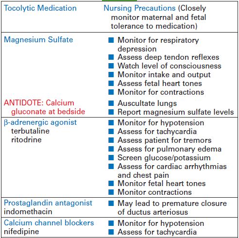
  11. Initiate tocolytic therapy


Pregnancy Complications - 2


Vaginal Bleeding (after 20 weeks’ gestation)

The complications may be related to placenta previa or abruptio placentae.

Placenta Previa
It is a low-lying position of placenta in the uterus that partially or completely covers the cervical os. Clinical Findings:
  • Painless bright red vaginal bleeding
  • Bleeding may be reported after intercourse
  • Uterine tone soft upon palpation
  • Interventions dependent on amount of bleeding and labor status
  • If partial placenta previa is noted in early gestation, then repeat ultrasound later in pregnancy (may demonstrate absence of previa as uterus grows)
  • If labor active and os is covered, then cesarean birth necessary
  • If bleeding controlled and labor absent, then conservative management is applied

Patient Teaching (Conservative Management)
  • No tampon use
  • No sexual intercourse
  • Monitor and report bleeding
  • Patient instructed to report placenta placement when admitted to hospital
  • Cesarean preparation class
  • Count fetal movements


Abrupto Placentae

Clinical Findings
  • Abdominal pain (sudden onset, intense and localized)
  • Fundus firm, boardlike, with little relaxation
  • Vaginal bleeding
  • Bleeding may be concealed within the uterine cavity
  • Fetal heart tones may be non-reassuring
Nursing Care (vaginal bleeding/late pregnancy)
  1. Monitor amount of bleeding
  2. Check vital signs
  3. Observe for signs of shock
  4. Evaluate fetal heart tones
  5. Palpate uterine tone
  6. Apply electronic fetal monitor (EFM)
  7. REPORT alterations in fetal heart rate pattern
  8. REPORT hypertonic contractions with poor resting tone
  9. Do not attempt vaginal exam until placenta placement verified
  10. Initiate IV fluids
  11. Report laboratory and ultrasound findings
  12. Prepare staff for possible cesarean birth
  13. Attend to patient’s emotional needs


Pregnancy Complications - 1

Vaginal Bleeding (before 20 weeks’ gestation)
It may be related to spontaneous abortion, ectopic pregnancy, or gestational trophoblastic disease.

Spontaneous Abortion
It is a loss of pregnancy before viability, clinical Findings:

  • Vaginal spotting (may pass clots)
  • Abdominal cramping
  • Cervical changes
  • Fetal heartbeat may be present or absent

Ectopic Pregnancy
It is a product of conception implant outside the uterus, clinical findings:

  • Vaginal spotting
  • hCG lower than expected for dates
  • Lower abdominal pain
  • Ultrasound findings: absence of intrauterine gestational sac
  • If rupture occurs, it would be: Positive Cullen’s sign (periumbilical bluish hue), Shoulder pain, and Signs of shock

Gestational Trophoblastic Disease
It is an abnormal proliferation of trophoblastic cells without viable fetus, Clinical Findings:

  • Vaginal spotting (dark brown)
  • Fundal height greater than expected for dates
  • hCG greater than expected for dates
  • Excessive nausea and vomiting
  • Absence of fetal heart tones
  • Ultrasound findings: Snowflake-like clusters, absence of fetus

Nursing Care (vaginal bleeding/early pregnancy)

  1. Monitor amount of bleeding
  2. Assess vital signs
  3. Observe for signs of shock
  4. Auscultate for fetal heart tones (FHTs)
  5. Collect passed tissue/clots
  6. Monitor patient comfort
  7. Check blood type and Rh factor
  8. Administer Rh(D) immunoglobulin if indicated
  9. Initiate IV fluids as ordered
  10. Report lab/ultrasound findings
  11. Attend to patient’s emotional needs


Forceps Birth

Forceps birth is an assisted birth, sometimes called an instrumental or operative vaginal birth, uses instruments that are attached to baby’s head so that baby can be pulled out. Forceps are described as stainless steel that come in two intersecting parts and have curved end to cradle baby’s head. It can be used as low or outlet forceps or for mid-forceps procedures.
This procedure is done to provide traction or to assist in rotation of the fetus.

Advantages:
  • Provide assistance when laboring women is exhausted
  • May decrease need for cesarean birth
Disadvantages:
  • Maternal complication such as vaginal and perineal lacerations and postpartal hemorrhage
  • Neonatal complication such as facial bruising, edema and cerebral trauma.
Nursing care:
  • Explain the procedure to woman
  • Encourage her to relax perineum and breathe during forceps application
  • Advice physician when contraction is present
  • Assess newborn for facial bruising or edema.

Induction

Induction labor is a procedure to stimulate uterine contraction during pregnancy before labor begins spontaneously. It is done for various reason, especially for mother and baby health reason. Elective induction may be accomplished by oxytocin infusion.

Advantages:
  • IV oxytocin induction is usually successful when labor readiness has been established, fetal maturity is established and Bishop score is 9 or more.
  • Maternal and fetal status can be monitored closely.
Disadvantages:
Induction is an invasive procedure.
Hypertonic labor, fetal distress, alterations in blood pressure, ruptured uterus.

Indications:
  • Postmaturity
  • Premature rupture of membranes
  • PIH
  • Presence of maternal disease such as diabetes mellitus
  • Fetal demise.
Contraindications:
  • Grand multiparity
  • Placental abdominalities
  • Previous uterine surgery
  • Fetal distress
  • Preterm fetus
  • Positive CST
  • Abnormal fetal presentation
  • Presenting part above inlet
  • Cephalopelvic disproportion (CPD).
Nursing Intervention:
  • Obtain baseline tracing of uterine contractions
  • Follow established protocols
  • Increase IV dosage only after assessing contractions, FHR, and maternal blood pressure and pulse.
  • Do not increase rate once desired contraction pattern is obtained.
  • Discontinue oxytocin if contraction frequency is less than 2 minutes of duration is more than 90 seconds, or if fetal distress is noted.

Amniotomy

Amniotomy is the artificial rupture of membranes, sometimes called as AROM. It is done to stimulate labor.

Advantages:
  • Amniotomy can stimulate contractions
  • Amniotomy can evaluate the amniotic fluid
Disadvantages:
  • Birth must occur within 24 hour when amniotomy is done, and may be need cesarean birth.
  • Increased risk of prolapsed cord
  • Risk of Infection
Nursing Care:
  • Auscultate Fetal Heart Rate before and after amniotomy
  • Record the time of Amniotomy, Fetal Heart Rate, and characteristics of fluid (amount, color and odor).
  • Instruct woman to remain in bed unless fetal presentation part is well engaged. It is done to prevent prolapsed of umbilical cord.
Risks for Mother:
  • Increases the risk of infection.
  • Labor may become more aggressive
  • The mother increases her chances of having uneven dilation.
Risk for Baby:
  • Increase of umbilical cord compression
  • The pressure on the baby’s head causes swelling in some part.

Diabetes in Pregnancy

Client with diabetes and their infants are at risk for complication during pregnancy. Infants of diabetic mother tent to be large for gestational age. It is caused by glucose that crosses the placenta, whereas insulin does not, these infants tend to gain weight. The problem is that high glucose environment impedes lung development and although the infants are large for gestational age, they are often premature.

Complication of infants from maternal diabetes:
  • Patent ductus arteriosus (PDA)
  • Polyhydramnions
  • Premature delivery
  • Respiratory distress syndrome
Complications of mother with diabetes pregnancy:
  • Hypertension
  • Renal disease
  • Ketoacidosis
  • Vascular compromise
  • Seizure activity related to hypoglycemia
Another problem in diabetes pregnancies is that the fluctuations in maternal blood sugar can result in fetal brain damage or sudden fetal death due to ketosis. That's why the client should be taught to check their blood glucose levels frequently during the day. Level over 120 mg/dL should be reported to the doctor for treatment.

Infants born to diabetic mothers might be delivered by cesarean section because of their large sizes and they should be assessed immediately after delivery for hypoglycemia by performing a dextrostix. The glucose level of 40 mg/dL or lower indicates hypoglycemia in the infant.

The blood is usually drawn from a heel stick and should be stuck on the lateral aspect of the heel. Blood test should be performed to detect hypocalcemia, hypokalemia and acidosis status.

Ectopic Pregnancy

Ectopic pregnancy is the condition in which the ovum implants in area other than the endometrial lining of the uterus. This pregnancy is not commonly successful since the areas outside of the uterus cannot sustain for a full-term pregnancy. It’s studied that it usually happens when there is a tubal blockage that prevents the fertilized ovum from passing through the fallopian tubes.

Ectopic pregnancy can be happened at abdominal, tubal, myometrial or cervical.

Ectopic pregnancy at abdomen:
The abdomen is usually unable to sustain for embryo growth

Ectopic pregnancy at tubal:
This is the most common site of ectopic pregnancy. It can causes mother at risk for tubal rupture that can be a life threatening condition.

Ectopic pregnancy at myometrial:
We cannot recognize it until delivery that usually requires a hysterectomy to stop bleeding. Sometime it is called as placenta accrete.

Ectopic pregnancy at cervical:
It has relation with placenta previa

Precipitating Factors:
  1. Pelvic Inflammatory Disease
  2. Previous tubal surgery or tubal pregnancy
  3. Endometriosis, and
  4. Congenital anomalies of the fallopian tubes
Sign and Symptoms:
  1. Sharp one-sided pain
  2. Tenderness of adnexal, area over ovary and tube
  3. Vaginal bleeding (may or may not seen)
  4. Hard and rigid abdomen and signs of circulatory collapse when tubal is ruptured.
How to care patient with ectopic pregnancy:
  • Provide emotional support for whom undergoing surgical or medical treatment
  • Provide emergency resuscitation and emergency surgery
  • Teach mother about pre and post operative self care
  • Consider to refer mother to a Fetal Demise Support Group

Anesthesia in Labor and Delivery

There are five types of anesthesia used in labor and delivery: local anesthesia, pudendal block, lumbar epidural block, subarachnoid (spinal) block, and general anesthesia.

Local Anesthesia:
  • It is used for blocking pain during episiotomy
  • It is administered just before the birth of the baby
  • The anesthetic has no effect on fetus

Pudendal Block:
  • It is administered just before the birth of the baby
  • The anesthetic is injected into the pudental nerve through a transvaginal route
  • It has effect last about 30 minutes
  • It blocks the perineal area for episiotomy
  • There is no effect on contraction or the fetus


Lumbar Epidural Block:
  • The anesthetic relieves pain from contractions and numbs the vagina and perineum
  • The anesthetic is injected in epidural space at L3 to L4 and may cause hypotension, so assess the maternal blood pressure
  • The anesthetic is administered after labor is established or just before a scheduled casarean birth
  • Keep mother in side-lying position or place a rolled blanket beneath the right hip to displace the uterus from the vena cava
  • Administer IV fluids if prescribed
  • Increase fluids if hypotension occurs

Subarachnoid (spinal) Block:
  • The anesthetic is injected into the spinal subarachnoid space at L3 to L5 and administered just before the birth
  • It relieves uterine and perineal pain and numbs the vagina, perineum, and lower extremities
  • The anesthetic can cause maternal hypotension and postpartum headache
  • Keep mother lie flat for 8 to 12 hours following spinal injection
  • Administer IV fluids as prescribed

General Anesthesia:
  • General anesthesia may be used for some surgical interventions
  • It might cause a danger of respiratory depression and vomiting
  • The mother is not awake

STAGES OF LABOR – Stage IV

LAST STAGE

  • Last stage of labor begins with delivery of placenta and ends with postpartum stabilization
  • Duration: usually 1-2 hours after delivery (primipara or multipara)
  • Blood pressure returns to the pre-labor level
  • Pulse is slightly lower than during labor
  • Fundus remains contracted, in the midline, 1-2 fingerbeadths below the umbilicus

Nursing Interventions:
  • Maternal assessment every 15 minutes for 1 hours, every 30 minutes for 1 hours, and hourly for 2 hours
  • Administer oxytocin product if ordered
  • Assess fundus every 15 minutes, if soft, massage with side of hand
  • Assess lochia, checking peripad and under lower back
  • Assess bladder for distention because full bladder will prevent contractions and increase bleeding
  • Assess episiotomy for intactness and possible bleeding

STAGES OF LABOR – Stage III

THIRD STAGE
  • Third stage of labor begins with delivery of infant and ends with delivery of placenta
  • Duration: up to 20 minutes (primipara or multipara)
  • Contractions occur until the placenta is born
  • Placental separation and expulsion occur
  • Birth of placenta occurs 5-30 minutes after birth the baby

Nursing Interventions:
  • Assess maternal signs and uterine status
  • Observe for placental separation
  • Observe mother for signs of altered LOC or altered respiration (indicate aneurysm or emboli)
  • Allow maternal-infant interaction as soon as possible

STAGES OF LABOR – Stage II

SECOND STAGE


  • Second stage of labor begins with complete dilation and ends with delivery of infant
  • Duration: 30-90 minutes in primipara and 15-20 minutes in multipara
  • Cervical dilation complete
  • Uterine contractions occur every 2-3 minutes, lasting 60-75 seconds and the intensity is strong
  • Increase in bloody show
  • Mother feels urge to bear down

Nursing Interventions:
  • Assess fetal well-being continuously
  • Monitor maternal vital signs
  • Encourage pushing
  • Encourage deep-full breath (not to hold breath longer than 5 seconds when pushing)
  • Commend mother’s effort

STAGES OF LABOR – Stage I

First Stage

The first stage consists of three phases: latent, active, and transition. This stage begins with the first true contraction and ends with complete effacement and dilation to 10 cm.

Latent Phase (Early Labor):
  • Duration: 10-12 hours in primipara and 8-10 hours in multipara
  • Cervical dilation is 1 to 4 cm
  • Uterine contractions occur every 15-30 minutes and are 15-30 seconds in duration and mild intensity
  • Mother is talk active
  • Encourage mother and partner to participate in care
  • Change position and ambulation to comfort mother
  • Offer fluids an ice chips
  • Inform the progress to mother and partner
  • Encourage voiding every 1-2 hours

Active Phase:
  • Duration: 2-4 hours in primipara and 2-4 in multipara
  • Cervical dilation is 4-7 cm
  • Uterine contractions occur every 3-5 minutes and are 30-60 seconds in duration and of moderate intensity
  • Mother becomes restless and anxious as contractions become stronger
  • Mother may experience feeling of helplessness
  • Encourage mother in maintenance of effective breathing
  • Provide a quiet environment
  • Inform the progress to mother and partner
  • Backrubs, sacral pressure, pillow support and position changes to promote comfort
  • Offer fluids and ice chips
  • Instruct partner in effleurage
  • Encourage voiding every 1-2 hours

Transition Phase:

  • Duration: 2-4 hours in primipara and 1-2 in multipara
  • Cervical dilation is 8-10 cm
  • Uterine contractions occur every 2-3 minutes and are 45-90 seconds in duration and strong intensity
  • Mother may becomes tired, restless, irritable, and feels out of control
  • Encourage rest between contraction
  • Inform the progress to mother and partner
  • Provide privacy
  • Offer fluids and ice chips
  • Encourage voiding every 1-2 hours


Special Nursing Interventions First Stage:
  • Monitor vital signs
  • Monitor fetal heart rate via ultrasound Doppler, fetoscope or electronic fetal monitor
  • Assess fetal heart rate before, during and after a contraction (normal FHR is 120-160 beats per minute)
  • Monitor uterine contractions by palpating, determining frequency, duration, and intensity of contraction
  • Assess status of cervical dilation and effacement
  • Assess fetal station presentation and position by Leopold’s maneuver
  • Assess the color of the amniotic fluid if the membranes have ruptured because meconium-stained fluid can indicate fetal distress

Fetal Distress

Fetal distress is a compromise of the fetus during the antepartum period (before labor) or intrapartum period (birth process). It is commonly used to describe fetal hypoxia (low oxygen levels in the fetus).

Fetal distress can be detected due to abnormal slowing of labor, the presence of meconium (dark green fecal material from the fetus) or other abnormal substances in the amniotic fluid, or via fetal monitoring with an electronic device showing a fetal scalp pH of less than 7.2


Signs and Symptoms of Fetal Distress:

Nursing Interventions:

  • Place patient in a lateral position, elevate legs
  • Administer oxygen at 8-10 L/min via face mask
  • Discontinue oxytocin (Pitocin) if infusing
  • Monitor maternal and fetal status
  • Prepare for emergency cesarean section

Supine Hypotensive Syndrome

Supine hypotensive syndrome occurs when the venous return to the heart is impaired by the weight of the uterus. It results in partial occlusion of the vena cava and descending aorta and in reduced cardiac return, cardiac otuput, and blood pressure

Signs and Symptoms:
  • Hypotension
  • Fetal distress
  • Faintness, light-headedness, dizziness


Nursing Interventions:
  • Place patient in a lateral recumbent position
  • Monitor vital signs and fetal heart rate
  • Treat for shock if other signs of shock are present

Amniotic Fluid Embolism

Amniotic fluid embolism is the condition in which the amniotic fluid is escaped into the maternal circulation. It is usually fatal to the mother because the debris containing amniotic fluid deposits in the pulmonary arterioles.

Signs and Symptoms:
  • Respiratory distress and chest pain
  • Seizures
  • Cyanosis
  • Heart failure and pulmonary edema
  • Fetal bradycardia and distress

Nursing Interventions:
  • Emergency action is performed to maintain life
  • Administer oxygen at 8-10 L/min by face mask or resuscitation bag
  • Position patient on side
  • Prepare for intubation and mechanical ventilation
  • Administer IV fluids, blood products, and coagulation therapy
  • Monitor fetal status
  • Prepare for emergency delivery
  • Provide emotional support for patient, partner and family

Uterine Inversion

Uterine inversion is a condition that the uterus turns inside out completely or partly. It usually occurs during delivery or after delivery of placenta.

Signs and Symptoms of Uterine Inversion:
  • Severe pain
  • Hemorrhage
  • Depression in the fundal area
  • Interior of the uterus may be seen through the cervix or protruding the vagina

Nursing Interventions:
  • Monitor for signs of hemorrhage and shock and treat shock
  • Prepare patient to reposition the uterus to the correct position via the vagina or laparatomy if unsuccessful

Placenta Abruptio


Placenta abruptio is premature separation of placenta from the uterine wall after 20 weeks of gestation and before the fetus is delivered.

Signs and Symptoms:
  • Painful vaginal bleeding (dark red)
  • Uterine rigidity and tenderness
  • Severe abdominal pain
  • Signs of maternal shock
  • Signs of fetal distress

Nursing Interventions:
  • Monitor maternal vital signs and fetal heart rate
  • Assess for excessive vaginal bleeding, abdominal pain, and increase in fundal height
  • Bed rest, oxygen, IV fluids, and blood products as prescribed
  • Monitor and report any uterine activity
  • Prepare for the delivery of the fetus as quickly as possible
  • Monitor for sings of disseminated intravascular coagulation in the postpartum period
  • Administer Rh immune globulin if the mother is Rh-negative and has not been given the injection at 28 weeks of gestation